Today's discussion will go through a different dimension and for sure, it's something you are familiar with. What I intend to achieve here is to bring to you in the easiest way to understand, the heart function and physiology. How it operate independently without requiring electrical signal supply from other organs and tissues.
It is no news that the heart is the major organ responsible for pumping and distribution of oxygenated blood throughout the entire body system, both in humans and other animals. The pumping action of the heart is very significant for the continuity of life. Tissues and organs that are deprived of oxygen supply by any means end up necrosing (pathological death of cells).
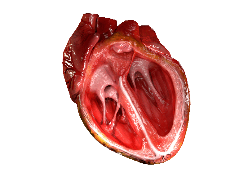
The human body is regarded as a system because, most of the organs function depending on the adequate supply of nutrients and oxygen. As long as the blood supply is adequate, the heart and other organs are expected to continue performing effectively at optimum rate. The main intent of this write-up, is to reveal those unsong features of the heart and our focus is on the independent attributes of the hearts as regarding electricity generation.
Why does the heart continue its beating action even after removed from the body?
One of the remarkable feature of the heart is its ability to continue its pumping/beating function despite being removed from the body system. I only got to understand and know this during my med school days. That being said, let's get a good grasps and understanding of this amazing feature of the heart.
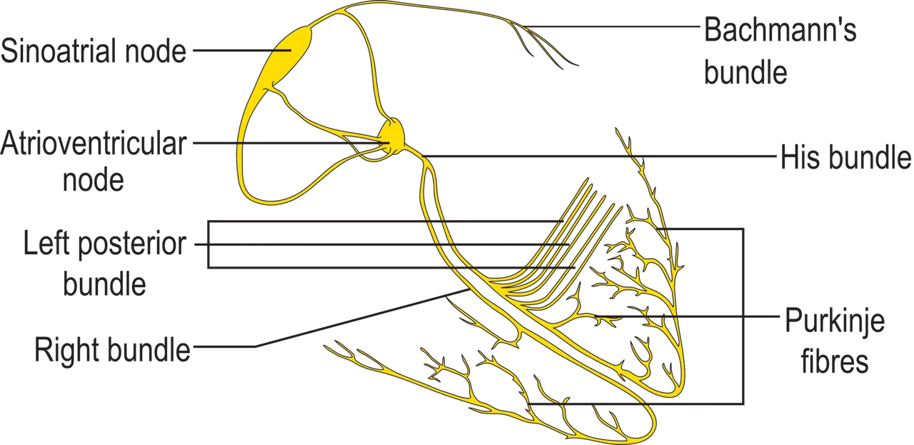
Without wasting time, let's go straight and answer the question. The heart is made up of what is known as the Sinoatrial node aka (SA node). This structure is a bundle of nerve that is responsible for the heart beat. It is the sole structure that is responsible for the beating action of the heart. It generates the electricity needed to power the pumping action of the heart. In a nutshell, it is aka the pace maker.
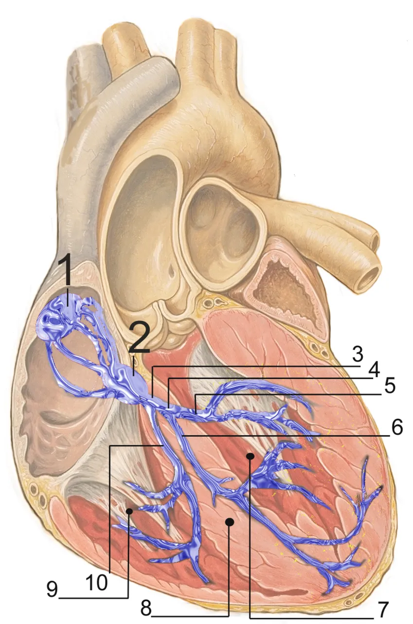
(1) Sinoatrial node, (2) Atrioventricular node, (3) Bundle of His, (4) Left bundle branch (5) Left (6) posterior fascicle (7) Left - anterior fascicle, (8) Left ventricle, (9) Ventricular septum, (10) Right ventricle, (10) Right bundle branch
When the heart is removed, probably in the process of heart transplant, it can still survive for about 3-4 hours before it runs out of the energy powering it (if all things being equal). We will explain this better as we go on. The current generated by the heart is from the SA node while the energy of contraction and relaxation come from those stored in the cardiac muscles cells aka the myocytes.
Cells in organs and tissues need nutrients, energy and most important oxygen to continue their functions, when they are deprived of this basic need, they could die. The heart is no different from other organs in this regards. In order to preserve this energy source, they are immediately put in a confinement that contains ice. What the ice does is to temporarily halt the beating action, cellular and metabolic activities going on in the heart, so that it doesn't run out of energy reserve.
So long as the heart is placed in the ice, traditionally, it can stay for as long as 3-4 hours. This traditional method has long been replaced with a more advanced technology and the sole aim of this technology is to increase the duration the heart can last outside the body, as well as maintain the heart in its normal physiologic state. This technology is called Heart in a box
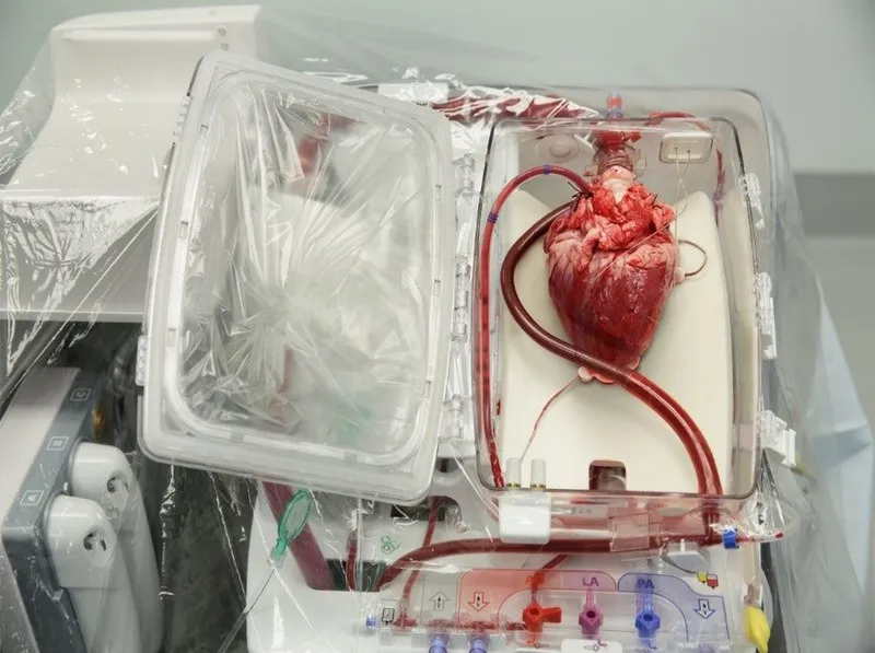
The major difference between this advanced technology and the traditional ice technology is that, warm oxygenated blood of the donor is continuously passed through the heart muscles constantly while it is outside of the body. This way, the heart can continue to beat indefinitely until it is ready to be transplanted into the recipient.
Heart transplantation even though a very good approach, still has its ugly sides. The possibility of rejection can come up later and that's why most patients that have undergone organ transplant in general are usually given immunosuppressant drugs to reduce the chances of auto immune destruction of self cells. Even though efforts are made to reduce this from happening, the possibility can for sure never fade.
What really is behind the activity of the SA node
The SA node is located just at the crista terminalis junction that is found around the right atrium's upper wall and also more specifically at the opening of the superior vena cava. It is at this point it generates and sends the electrical impulse that sets the heart rhythm. The action of the sinoatrial node is controlled by the autonomic nervous system.
Looking at the SA node, it takes the shape of a crescent and extends down, branching to other parts of the cardiac muscle. It is mostly made up of different myocytes that are separated by a connective tissue. These myocytes are the major cells that generate the electric impulse we have been talking about.
When we are at rest, the normal heart rate is within the range of 60-100 beats per minute. But during exercise or activities, it rises. You may be wondering why there has to be a range since the impulse comes from just a structure. Truth is that, the SA node is made up of multiple myocytes, and these myocytes generate different electric signals that vary from each other.
The reason for the slight variation in the rate of heart beat is simply because of different myocytes with different electric signal generation time. The rate at which they produce electric signal vary, even though these signals are still within the normal range. Since they produce the signal at varying time, the heart beats will always fluctuate. What this implies is that, you cannot expect to have the same values for cardiac rate when you measure it.
It is important we understand that, the conduction of the electric impulse generated by the SA node has to be transmitted to parts of the cardiac muscle. For contraction to occur, the electric impulse generated by the SA node must be transmitted to the Atrial ventricular node (AV node). The atrial node is the major distributor and transmitter of the electric signal generated by the SA node.
The AV node is located in an anatomical position know as the Koch triangle. The major function of the AV node is to transmit the signal from the atrium to the ventricle located just below the atrium.
The purpose of this structure is to connect the electrical systems of the atria and the ventricles, providing electrical impedance from the atria and an intrinsic pacemaker in its absence.
From the AV node, the signal is sent through the bundle of HIS. This structure then disperses this signal to various parts of the ventricles and even the left atrium. The efficiency of the bundle of HIS is very important to ensure synchronous heart beat. Bundle of HIS is the major structure that supplies the rest of the heart. A block in this structure causes the heart rate to drop and when this happens, it would require the need of a pace maker. The common cause of bundle block is the presence of blood clot in the lung, cardiomyopathy, chronic obstructive pulmonary disorder e.t.c.
In a nutshell, the SA node is solely responsible for generating the electric current that powers the heart even when the heart is removed from the ribcage aka sternum; where it is heavily protected against any form of injury or undue pressure exertion. Any form of disease or challenge that is associated with the SA node can result to what is regarded as Sinus node dysfunction.
Note at this point that, blockage in Bundle of HIS is not the only condition that may warrant the use of a pace a maker. In sinus node dysfunction, what you have is irregular rhythmic contraction of the heart. Sinus node dysfunction can warrant the use of an artificial pace maker. So what is an artificial pace maker?
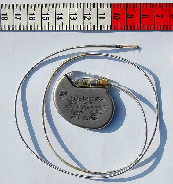
An artificial pace maker as the name implies, is simply an artificially produced device that is designed to mimick the functions of the SA node by independently producing electric impulse require for the metabolic activities of the heart such as contractions and relaxation of the cardiac muscles.
This device not only helps stimulate the pumping action of the heart, it is mainly used to monitor the heart rhythm and its rate. The ability of this device to monitor and know when the heart beat is abnormal, makes it an incredible innovation in the field of biomedical engineering and medicine. This device fills the gap created by some disease conditions that make the heart underperform.
To understand the heart functions and performance, it is easily interpreted as waves which is referred to as an electrocardiogram. The ability to differentiate between a normal and abnormal heart electrocardiogram helps us properly diagnose disease conditions associated with the heart, case in point, state of the electrical conduction system.
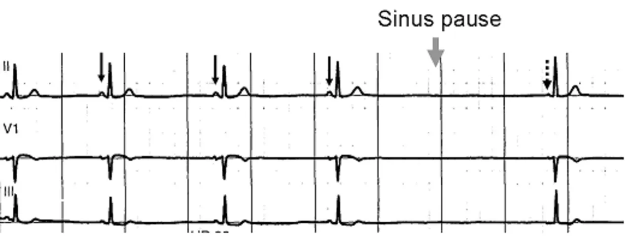
Normal sinus rhythm, with solid black arrows pointing to normal P waves representative of normal sinus node function, followed by a pause in sinus node activity (resulting in a transient loss of heart beats). Note that the P wave that disrupts the pause (indicated by the dashed arrow) does not look like the previous (normal) P waves- this last P wave is arising from a different part of the atrium, representing an escape rhythm.
It takes years of consistent experience and practice to have a good grasps of electrocardiogram readings. Needless to say that accurate electrocardiogram results play significant role in diagnosis and treatment of cardiovascular diseases. A wrong interpretation can lead to a wrong treatment approach. Let's round up our discussion by bringing to light some scientifically proven facts about the heart you probably don't know.
Undeniable facts about the heart
The first on our list is about exercise. No doubt, exercise has been proven to be a very important activity one must engage in to ensure good health. The relationship between the heart and exercise lies in the concentration of a type of inflammatory protein known as C-reactive protein.
Exercises helps reduce the concentration of this protein in the blood and thus reduces the risk of cardiovascular diseases. When you are actively engaged in exercise, it does not only reduce the concentration of the C-reactive proteins, but also reduces the level of low density cholesterol that cause atherosclerotic plagues in the heart coronary arteries. The coronary artery is just about 4mm in diameter. So you can imagine what will happen if there is any form of obstruction (e.g fats) to this vessels.
You can read more about this from my previous here - Do waste trainers have any positive effects on weight reduction in women. Reading the article will help you gain a better knowledge about cholesterol and their association with cardiovascular diseases.
Another interesting fact about the heart is the number of times it beats per day. Report has shown that the heart beat 100,000 times per day. Multiplying this value by 365 days in a year, would give a rough estimate of about 36500000 times per year. If we move this value further to know the number of times it beats in an average adult age of 70years, then we would have 2,555,000,000 (2.5 billion) times. This shows the remarkable work the heart does while it pumps s total of about 1.5 gallons of blood per day.
Another interesting fact about the heart is that, it is rare to have heart cancer. You are free to ask how is this? The reason is that, the cells of the heart stop dividing at the very early stage of life. This inability is a limiting factor for the progression of any cancer. Cancer cells take advantage of the replicative ability of cells to divide. Since the heart cells do not divide at latter stage life, it is hence, almost impossible for someone to have heart cancer.
If you think the above is all, you would probably be wrong. The heart is the organ that supplies all organs, tissues and cells in the body but unfortunately, there is a particular structure's cells in the body that does not receive blood from the heart. This structure is the cornea. The cells in cornea of the eyes does not receive any blood supply from the heart rather it only receives nutrients through diffusion from tear fluid
The heart is usually located on the left side of the body but in some cases and in individuals, it can be located on the right side of the body. Similarly, the apex of the heart could point to the right as against the normal position, which is the left.
Conclusively, the heart is a very important organ that is very crucial for life. Cardiovascular diseases can cause the heart to under-perform. Exercise is the cheapest therapy to maintaining a good heart. Cardiovascular diseases have high mortality and morbidity rate and more common in the aged.
References
•The Sinoatrial Node Function
•Sinoatrial Node Structure, Mechanics, Electrophysiology and the Chronotropic Response to Stretch in Rabbit and Mouse
•Evidence of Superior and Inferior Sinoatrial Nodes in the Mammalian Heart
•The sinoatrial node, a heterogeneous pacemaker structure
•5 Heart Facts That May Surprise You
•What Is the Heart? Anatomy, Function, Pathophysiology, and Misconceptions